canine eye diseases

What is Retinal Detachment?

blog_ Retinal Detachment _banner.png

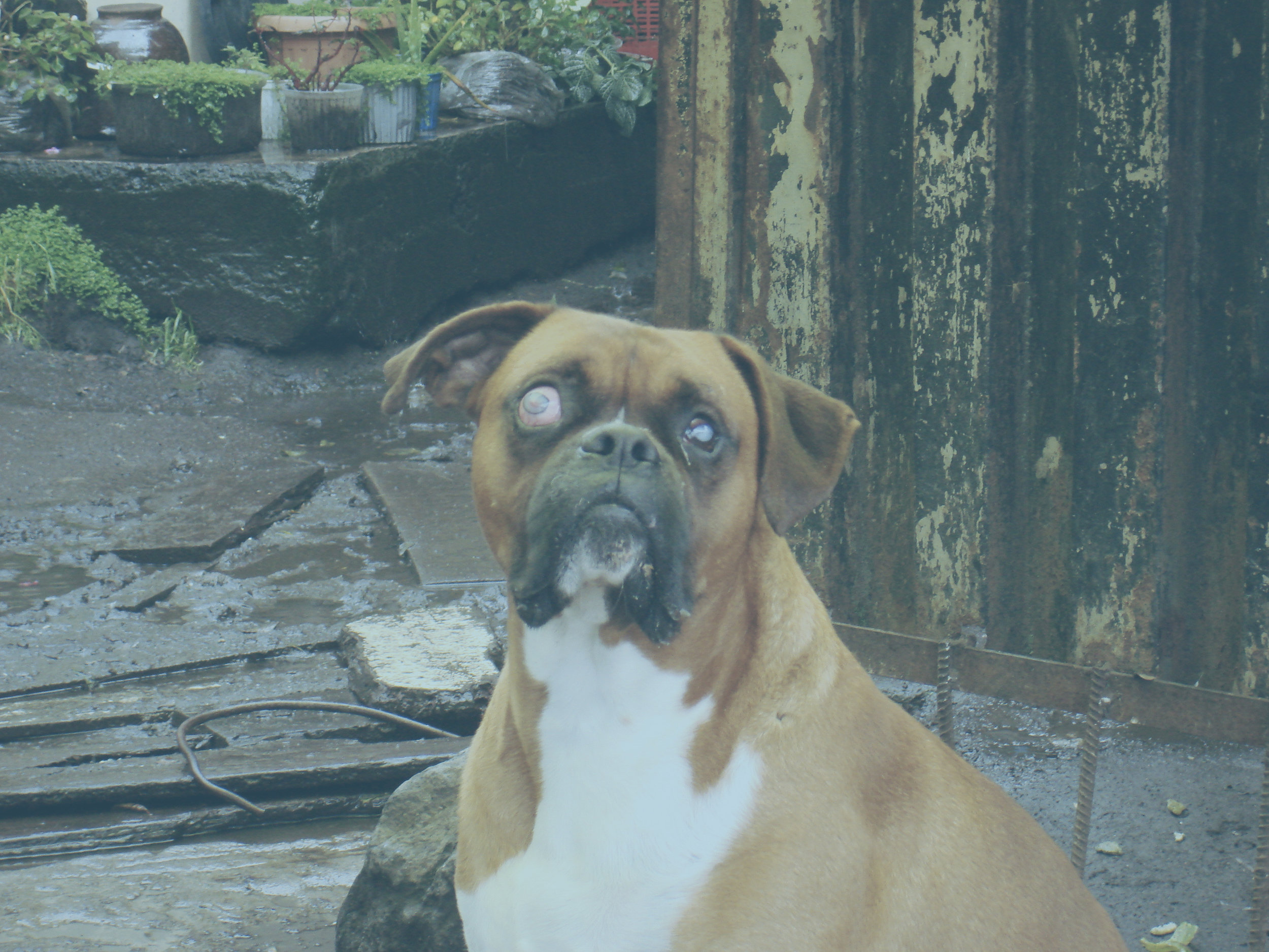
The retina is the light-sensitive tissue that lines the inner surface of the eye. When it becomes detached from the tissue supporting it, a very serious situation exists. It is extremely important to get your pet to the veterinarian immediately if you suspect they are having vision problems.


There are several factors that can cause this disorder.

Some of the most common factors include:

  • Injuries to the face or eye

  • Diabetes

  • Tumors

  • Infections

  • Kidney disease

  • High blood pressure (especially in cats)

  • Hyperthyroidism (in cats)

  • Sickle-cell anemia

  • Poisoning

  • Cataracts or cataract surgery

  • Genetics

  • Poor blood clotting

Symptoms

The most serious symptom of a retinal detachment is reduced vision or, in some instances, blindness. The severity of your pet’s ability to see is directly related to the seriousness of how detached her retina is, or if it impacts both eyes. Other symptoms include dilated pupils (when the eye shows no response to changes in light), discoloration of the white of the eye, or leaking of the eye and clumsiness due to your pet’s inability to see well.

Diagnosis

Your veterinarian will perform a complete history and physical examination, including a thorough ophthalmic examination. They may also refer you to a veterinary ophthalmologist for additional evaluation. In addition to providing a thorough examination of your pet, your veterinarian may recommend tests to identify the underlying cause.

These tests may include:

  • Chemistry tests to evaluate kidney, liver, and pancreatic function, as well as sugar levels

  • A complete blood count to screen for infection, inflammation, anemia, and other blood-related conditions

  • Electrolyte tests to ensure your pet isn’t suffering from an electrolyte imbalance

  • Screening tests to rule out infectious disease

  • Cultures, PCR testing, and other specialized tests, which can identify if specific parasites or diseases could be the cause

  • X-rays of the chest and abdomen to look for abnormalities

  • A fecal test to rule out fecal parasites

  • A thyroid test to determine if the thyroid gland is producing too little (in dogs) or too much (in cats) thyroid hormone.

  • Blood pressure measurement

Treatment

It is important to begin treatment as soon as possible to prevent further damage to the eye or permanent blindness. Treatment can include medications and/or surgery. It will depend on the underlying cause of the detachment, the severity of the condition, and your pet’s overall health.

Prevention

While you may not be able to prevent retinal detachment, by being a diligent pet owner and carefully checking your furry friend regularly for anything out of place, you will help catch problems in their earliest stages!

If you have any questions or concerns, you should always visit or call your veterinarian – they are your best resource to ensure the health and well-being of your pets.


Hear From Us Again

Don't forget to subscribe to our email newsletter for more resources and latest news delivered to your inbox (here). Or, you can keep up to date by liking and following our Facebook page (here).

Related: We have more information under our dog health and cat health tags.

Common Eye Problems in Dogs

Did you know a dog’s eye can develop more than just cataracs of the eye? Eye conditions have a tendency to worsen progressively, so it is important to recognize and discuss any concerns that you might have about your dog’s eyes with your veterinarian as quickly as possible.


1-cherry-eye.jpg

Eyelid Protrusion (Cherry Eye)

A dog has three eyelids. Two are readily visible and an extra one, called the third eyelid, normally hides from view just below the inner corner of the eye. The third eyelid is home to the tear producing gland that is normally hidden. However, some dogs have a congenital weakness of the ligaments that hold it in place. When the ligaments are weakened, the gland pops out of its normal location and looks much like a “cherry” at the inner corner of the eye. Due to it being a condition is due to genetics, both eyes are often affected over time. Treatment of cherry eye is simple— a veterinarian will perform simple surgery to attach the gland back to normal position.

2-corneal-wound.jpg

Corneal Wounds

The surface of the eye is covered by the cornea, which is a clear, skin-like tissue. Much like skin, the cornea can be injured. Lacerations (cuts), punctures, and ulcers are all quite common in dogs. Trauma to the eye is often to blame, like when a dog runs through tall grass and is poked in the eye. In other cases, other problems such as poor tear production or abnormal anatomy can put a dog at risk for corneal damage. A dog with a corneal wound will rub the affected eye and squint due to pain. The eye may also appear red and have excessive drainage. Treatment involves prevention or treatment of infections with antibiotic eye drops or ointments, managing pain, or simply giving the cornea time to heal. In severe cases of corneal wounds, surgery or other treatments may be needed to protect or repair the cornea.

Keratoconjunctivitis Sicca (KCS) or Dry Eye

When a dog develops a disease called keratoconjunctivitis sicca (KCS) or dry eye, the tear glands produce fewer tears than what is normal. Tears are important because they remove potentially damaging material from the surface of the eye and nourish corneal tissues. Believe it or not, a lack of tears can cause bigger problems, such as corneal ulcers, chronic drainage of mucus from the eyes, and pain. Treatment in mild cases of KCS can often be managed with frequent application of an artificial tear solution, but medications that stimulate tear production (i.e. cyclosporine) are usually necessary. In severe cases, one option is to perform a surgery that redirects a duct carrying saliva to moisten the eye.

Conjunctivitis (Pink Eye)

The conjunctiva are the mucus membranes that cover the inside of a dog’s eyelids, both sides of the third eyelid and some parts of the eyeball itself. Conjunctivitis and Pink Eye are interchangeable terms that simply mean inflammation of the conjunctiva. The symptoms of conjunctivitis include reddened and swollen conjunctiva, eye drainage, and discomfort.

Conjunctivitis is more of a symptom of a disease, not a disease itself. Many conditions in dogs cause conjunctivitis, including physical irritation (like dust or inward growing eyelashes), infections (bacterial and viral) and allergic reactions.

Treatment depends on the underlying cause. Sterile saline eye washes can be used to flush irritants from the eye. Bacterial eye infections are usually resolved when treated with the appropriate prescribed antibiotic eye drop or ointment. The chances of catching pink eye from your dog is very low, but it makes perfect sense to wash your hands thorough after applying your dog’s eye medications. Make an appointment with your vet if your dog’s conjunctivitis worsens or fails to resolve over the course of a day or two.

5-glaucoma.jpg

Glaucoma

Production and drainage of fluid is balanced with precision to maintain constant pressure in the eye. Glaucoma occurs when this balance is disrupted and pressure increases. Symptoms include pain, eye redness, increased tear production, a visible third eyelid, corneal cloudiness, dilated pupils, and in advanced cases, an enlarged eye.

Treatment may involve a combination of topical or oral medications that decrease inflammation, absorb fluid from the eye, lower fluid production within the eye and promote drainage of fluid from the eye. In some cases, surgery is also a considerable option. Do not delay in contacting your veterinarian if you are worried your dog may have glaucoma, as it can result in blindness.

6-entropion.jpg

Entropion

Eyelids that roll inwards is what is called entropion. Entropion causes hair to rub on the surface of the eye, resulting in pain, increased tear production, and eventually damage to the cornea. Entropion can develop as a result of chronic squinting due to discomfort or eyelid scarring. It can also be a congenital problem. A veterinarian can temporarily suture the eyelids to a more normal position (eyelid tacking) if entropion has occurred in result of a recoverable condition. Otherwise, there is surgery available to permanently fix the eyelid.

Progressive Retinal Atrophy (PRA)

Progressive retinal atrophy (PRA) can be hard to spot. It is a condition that causes dogs to gradually become blind over time while their eyes remain to appear normal. The first noticeable symptom of PRA is with difficulty seeing at night. Unfortunately, no effective treatment exists for PRA, but the condition is painless and dogs generally adapt extremely well to becoming blind.

8-cataracts.jpg

Cataracts

Cataracts block light from reaching the back of the eye, resulting in poor vision or blindness, depending on severity. Cataracts are often confused with lenticular sclerosis—a normal change for aging dogs that affects a dog’s lenses. Both conditions give the black center of the eye (pupils) a white, grey, or milky appearance. However, one can tell with a standard eye exam performed by your veterinarian. Cataract surgery is available when a dog’s vision is severely compromised, otherwise most dogs adapt very well to ailing vision.


Hear From Us Again

Don't forget to subscribe to our email newsletter for more recipes, articles, and clinic updates delivered to your inbox (here). Or, you can keep up to date by liking and following our Facebook page (here). We also have additional helpful articles under our tips category (here).