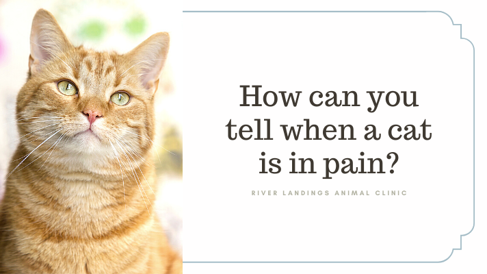
How can you tell when a cat is in pain?

Assessing pain is a complicated challenge, especially in cats. Pain has two primary components: the sensory aspect (intensity, location and duration) and the affective aspect (emotional toll).

Because pain assessment is somewhat subjective, veterinarians constantly try to create tools that make this process more objective. For validity, any pain measuring tool should take into consideration both characteristics: the sensory and the affective.

Signs of pain in cats

A British study was recently conducted in order to reach a consensus about criteria when evaluating pain in cats. Ultimately, 25 signs were considered to be reliable and sensitive for indicating pain in cats, across a range of different clinical conditions:

Top 5 Signs:

  • Appetite decrease

  • Avoiding bright areas

  • Growling

  • Groaning

  • Eyes closed

Other signs included: Lameness, difficulty jumping, abnormal gait, reluctant to move, reaction to touch, withdrawing/hiding, absence of grooming, playing less, overall activity decrease, less rubbing toward people, general mood, temperament, hunched up posture, shifting of weight, licking a particular body region, lower head posture, eyelids tightly shut, change in form of feeding behavior, straining to urinate, tail flicking

The top 5 signs are indicative of severe pain. Behavioral changes, such as irritability, tend to be seen with more long-term pain. The other signs can be observed with less intense pain. All of these signs cover both the sensorial and the emotional aspects of pain.

What if you see these signs of pain in your cat?

Cat owners should be aware of these signs. It is easy to mistakenly attribute behavioral changes, such as absence of grooming or playing less, as signs of aging; they can actually be signs of pain.

Remember, the presence of any single one of these 25 signs means pain. If you see any of these signs in your cat, see your veterinarian right away. Also remember that the absence of a sign does not mean your cat is no pain.

These signs may help both vets and cat guardians better assess the pain status of cats in their care.

While it can be fairly easy to recognize severe pain, it is much more difficult to detect low grade pain. The criteria above are a great start. Hopefully, this research will spark more studies to help us assess mild pain in cats as well to ensure their well-being.

If you have any questions or concerns, you should always visit or call your veterinarian— they are your best resource to ensure the health and well-being of your pets.


Hear From Us Again

Don't forget to subscribe to our email newsletter for more recipes, articles, and clinic updates delivered straight to your e-mail inbox.

Related Categories:

cat health

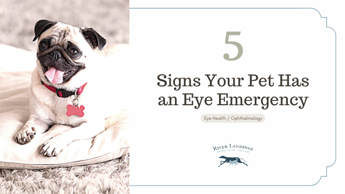
5 Signs Your Pet Has an Eye Emergency

5 Signs Your Pet Has an Eye Emergency

It's not uncommon for a veterinary clinic to field plenty of questions concerning pet eye problems on the daily. “Watchful waiting” is advised with some minor complaints, but never with eyes.

A non-vet pet owner cannot assess an eye problem’s severity, and a description over the phone just doesn’t cut it. In other words, when someone calls in and says their pet’s eye “looks funny,” it’s time for a trip to the vet.

Your vet may even direct you to a board-certified veterinary ophthalmologist for immediate assessment if the emergency requires it.

Red, inflamed or swollen eyes? Each of these symptoms can be caused by different ailments.

1. Red Eye

The white of the eye should be pristine white, with the odd lazy blood vessel meandering across the surface. If you’re not sure what this looks like, check out your own eye in the mirror.

Red eyes are not normal.

Gently lift the upper eyelid to check, and you’ll see anything from a rosy pink to a livid red. As a rule of thumb, the angrier the eye looks, the more urgently it needs checking.

Causes of red-eye range from irritation and infection to a condition called glaucoma.

Glaucoma: Pressure builds within the eye, like blowing too much air into a balloon. The most common cause is a problem within the eye and is often breed-related (Basset Hounds, Cocker Spaniels, terriers and northern breeds are at greatest risk). The pressure can damage the retina and cause blindness, so swift action is essential.

Conjunctivitis: Infection causes reddening of the eye. Sometimes the problem can be self-limiting, but — especially if there is a sticky yellow-green discharge, too — please see a vet.

2. Yellow-Green Discharge

It’s normal to have a white gloop in the corner of an eye first thing in the morning. Just wipe this away with a clean, damp cotton towel.

Also, rust-colored gloop is fine. This is a normal gloop that’s been exposed to the air for a while and oxidized (like when you cut an apple in half and it goes brown).

What isn’t normal is a thick, yellow-green discharge from one or both eyes. This is commonly a sign of infection.

Have the vet check the eye because some infections need antibiotics, while others can occur as a complication of another problem that needs attention.

3. Swollen Eye

If there’s something odd about your pet’s face, compare one eyelid with the other to see if one side is swollen. Eyelid swelling can be the result of an allergy, trauma, or infection. It’s best to seek vet attention because the eyeball needs checking to make sure it wasn’t damaged.

4. Dull Eye

Is the eye lackluster? A normal eye is bright, and you can see reflections on the surface— but sometimes the surface is dull and reflections aren’t clear, or those images are broken up or haphazard.

The most common reasons for this are either a dry eye or a corneal ulcer:

Dry eye: Our eyes are kept comfortable by the production of tear fluid. A pet with dry-eye fails to produce enough tear fluid, which leads to the surface drying out. One consequence is a dull surface, and another is the eye tries to protect itself by producing a thick, glue-like discharge. In the long term, scar tissue forms, impairing the vision.

Corneal ulcer: This is like a burst blister on the surface of the eye. In some cases, it heals on its own, but other times it can be dangerous and cause perforation of the eye.

5. Closed Eye

A closed eye is painful: Just think of the last time you had grit in your eye.

The pain might be due to a corneal ulcer, a knock to the eye, or a foreign body — anything from dust to grass particles or even a twig. When our pets are sniffing around, it’s not uncommon to get something lodged behind the eyelids.

Your vet will put drops of local anesthetic into the eye to get a better look and remove the object.


5 Most Common Eye Emergencies (in Dogs)

Here, we go into more detail about some of the top ophthalmologic emergencies we see with dogs in general practice.

1. Corneal Ulcers

Corneal ulcers are the most common acute eye problems seen. (This goes for cats as well.) The cornea is the outer “skin” of the eye; an ulcer occurs if this layer has been damaged.

Corneal ulcers are often traumatic in origin, although certain diseases of the cornea can result in an ulcer. Trauma to the cornea can occur with a scratch from a bush, a stick or even another critter.

Treatment involves early diagnosis of the severity of the ulcer and the administration of appropriate eye medications.

More serious ulcers may require surgery, and frequent re-checks are needed to ensure the cornea is healing nicely. When a corneal ulcer goes from bad to worse, the cornea can actually rupture — this is a true emergency indeed.

2. Proptosis

Proptosis occurs when an eye literally bulges out of the socket and the eyelids entrap the globe. This occurs most frequently in brachycephalic breeds and is a true emergency.

Even with immediate care, the dog may lose the eye depending on the extent of trauma sustained by the extraocular muscles, nerves, and blood supply.

Many of these eyes can be replaced and vision saved in about 20% of dogs, but only if you act immediately.

3. Corneal Laceration

Corneal laceration occurs when there is a complete tear through the cornea. The most common offender from a cat claw.

A sharp object, like a stick, can also puncture the cornea. The dog is almost always holding the eye completely shut and is in significant pain. Again, immediate surgery may save the eye.

Get to the vet. There is a very small window of opportunity when it comes to repairing these eye injuries.

4. Lens Luxation

Lens luxation is over-represented in certain breeds like the Russell Terrier and other terriers. In these dogs, due to a genetic disorder, the lens can spontaneously luxate, or become dislocated. In other breeds, causes may vary — head trauma is one example. This is a difficult diagnosis and may require a visit to an ophthalmologist.

Compared with the normal eye, a lens luxation can look like a very dilated pupil or a blue or whitish eye. Removing the lens can save the eye and save the dog from pain and total blindness.

5. Acute Glaucoma

Acute glaucoma looks like a discolored, a “red” or an inflamed eye. There may be discharge and painful blinking (called blepharospasm).

Glaucoma occurs when, for whatever reason, the pressure in the eye elevates, leading to pain, secondary changes, and blindness. The condition is usually obvious in just one eye, but both eyes are at risk.

Again, this is over-represented in certain breeds like the Cocker Spaniel, Basset Hound, Shih Tzu, Great Dane, and northern breeds.
As with every other eye problem discussed here, acute glaucoma should be assessed and treated immediately. Both systemic (oral) and ophthalmic drugs are used. Get an opinion from a veterinary ophthalmologist to help you and your primary care vet manage the case.

Don’t mess around with the eyes. Ophthalmic problems need early diagnosis and treatment.


Hear From Us Again

Don't forget to subscribe to our email newsletter for more recipes, articles, and clinic updates delivered straight to your e-mail inbox.

Related Categories:
DOG HEALTH

CAT HEALTH

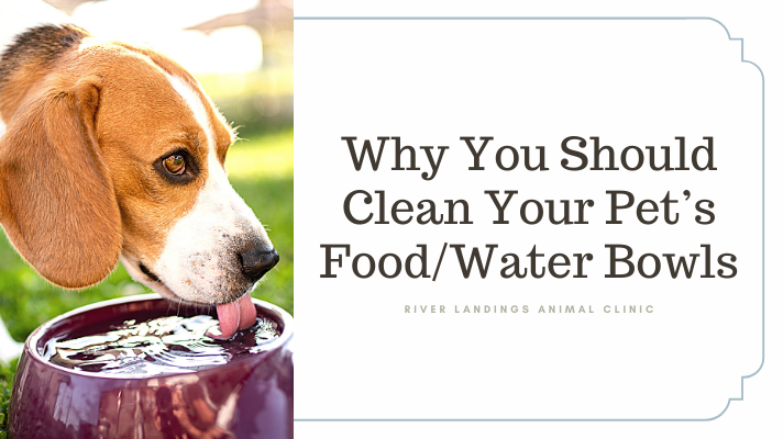
Why You Should Routinely Clean Your Pet’s Food and Water Bowls

Did you know your pet's food and water bowls are a hot spot of risk? Dirty food and water bowls can lead to parasites, affecting your pets and household. In the context of hygiene, pet food bowls are definitely an important part of your cleaning routine to be meticulous about.

We will cover a variety of things:

Good Hygiene & Safety Measures

4 Ways Your Dog’s Water Can Make Them Sick

Determining the right bowls for your pet

Where Do the Bugs Come From?

Zoonotic diseases are diseases that normally infect an animal, but poses a risk to people.

The most common zoonoses with regard to food and water bowls are salmonella, campylobacter, and worms.

These bugs have different mechanisms of causing infection, but they can all be shed into the environment through feces and travel indoors on the pet’s paws or coat.

When your pet licks their fur and then eats from a bowl, this can spread infection onto an object you touch. When you pick up the bowl, you run the risk of getting that same bug on your hands.

Also, with salmonella, the food itself may contain bacteria, with raw meat and eggs posing a special risk. We normally rely on thorough cooking to kill salmonella, so feeding a raw diet to your pet increases the risk.

Food Hygiene

Knowing raw meat could contain salmonella should make you think twice about where you store it.

It should be refrigerated (below 40° F) to slow down the growth of contaminating bacteria. Avoid storing raw meat or any pet food immediately beside human food. Make sure it’s securely wrapped and put in a different section of the fridge whenever possible.

The longer food sits in the open at room temperature, the greater the chance of bacterial contamination and multiplication. With this in mind, serve only the portion your pet can eat in one go so that food isn’t left sitting around. To streamline this and avoid the risks, buy packs of moist food in smaller sizes so you can throw unopened food away rather than store open sachets for days.

Related: Salmonella Infection in Pets & People and Zoonotic Diseases

What’s the Safest Way to Wash Bowls?

Wash them every day: Rinse them under running water and either hand-wash them in hot soapy water or put them on the top rack of the dishwasher (so the dishwasher’s heat doesn’t melt plastic dishes).

When hand-washing, do the pet’s dishes last, using a separate sponge or scourer kept just for the pet’s things. Make the water as hot as you can stand without burning yourself and use liquid or detergent soap. Rinse them well — your pets won’t enjoy a soapy aftertaste, and there’s a slim chance it could make them ill.

Fresh drinking water must be available for your pet at all times, so have at least 2 sets of everything so you have one bowl that’s clean and full of water for your pet while you are cleaning the other one.

Other Safety Measures

Always keep separate utensils for the pet’s food, such as serving spoons, forks, or scoops. Never use the pet’s things (toys, bowls, etc.) as a scoop.

Although some pets have nasty tummy upsets as a result of salmonella and campylobacter—not all do. Some pets are carriers, meaning the bug is in their system—they’re not ill, but they excrete it in their waste. Some of those bacteria (and also worm eggs) get onto the pet’s coat and then onto your hands when you stroke them. Delightful imagery, right? Wash! Your! Hands!


4 Ways Your Dog’s Water Can Make Them Sick

unsplash-image-pQVecS8pBNY.jpg

Your dog’s water bowl is the 3rd most germ-laden place in the house!

We all know clean and fresh water is vital for good health. With this in mind, you probably top up the bowl every time it goes down.

But as it happens, this isn’t sufficient to ensure good health. Merely topping up the water level does nothing to control harmful bacteria or even toxins that can be lurking in the water. Not only are these bugs unpleasant, but also they can cause illness in people, especially those with weak immune systems. So what bugs could be lurking in your pet's water bowl? Surprisingly, quite a few!

1. Giardia

This single-celled parasite may be better known to you as “traveler’s diarrhea.” It can infect people too—not just your pets.

Giardia is shed in feces inside a hardy oocyst. The latter is tough and can survive for long periods of time in warm, humid conditions, and thrives in water.

When a dog drinks infected water, the parasite passes into its gut. Some dogs show no symptoms, but others can develop sickness and weight loss in addition to diarrhea.

A diagnosis is made by examining a fecal sample under a microscope. Most cases respond well to 3-10 days of medication tailored to your pet.

Related: How do pets and people become infected with Giardia?

2. Cryptosporidium

Cryptosporidium is similar to giardia but a whole lot tougher. It’s so tough that it can survive chlorine treatment. Similar to giardia, oocysts are passed in feces that, if they get into the water, are a source of infection for animals and birds.

  • Your vet will prepare a treatment of fenbendazole or metronidazole to kill the parasite in the gut.

  • Bathe and shampoo the dog on the last day of treatment to wash away any oocysts clinging to the coat. Not doing so risks reinfection should the dog lick their fur.

Related: What Can I Catch From My Dog?

3. Leptospirosis

Leptospirosis is a particularly nasty infection with the potential to kill. It’s also a devious infection because signs can range from mild stomach upsets that last for weeks to months to an acute liver and kidney failure that are fatal in hours.

Other symptoms include:

  • Fever

  • Muscle weakness

  • Blood in the urine

  • Loss of appetite

  • Vomiting

  • Diarrhea

  • Collapse due to organ failure

Leptospirosis can be spread by rat urine and especially loves water sources such as standing water, puddles, and birdbaths. The unpredictable nature of leptospirosis means prevention is the best policy.

Luckily, there are vaccines available for your dog. If you live in a risk area for lepto, then your vet will advise giving the vaccine. A word of caution, though: The vaccine must be given yearly, as immunity is often gone completely 12 months after the previous dose.

Humans, however, don’t yet have a vaccine. Did you know pets can pass lepto to people? If your dog shows similar symptoms to those above and you feel unwell, see your physician as a matter of urgency.

Related: You and Your Pet Are at Risk For Leptospirosis

4. Blue-Green Algae

You may be aware that dogs shouldn’t swim in water with an obvious algal bloom—but do you know why?

Cyanobacteria, also known as blue-green algae, are a toxin-producing form of bacteria. This toxin can affect any mammal (yes, including people), causing nerve and liver damage.

Signs of toxicity include:

  • Weakness

  • Muscle tremors

  • Drooling

  • Vomiting

  • Diarrhea

  • Breathing difficulties

  • Collapse and death

There is no antidote to blue-green algae poisoning. Treatment includes rinsing out the stomach to remove contamination and supportive care with intravenous fluids.

So don’t let your dog swim in dirty water or drink from containers of “old” water — give them the fresh stuff.

Related: How Red Tide Affects Your Pet


Biofilms on Your Pet’s Water Bowl Are Bad

unsplash-image-yz4g1tYJti0.jpg

Spot the biofilm in your pet’s water bowl.

Does your dog or cat have a stainless steel drinking bowl? Next time you rinse the bowl under the tap, look for that misty, smoky layer covering the metal.

If you can’t see it, try wiping your finger across the wet metal surface. If your fingertip leaves a clean streak in its path, then you’re looking at a biofilm.

The definition of a biofilm is: “A thin but robust layer of mucilage adhering to a solid surface and containing a community of bacteria and other microorganisms.” So, basically a thin layer of stickiness rich in bacteria that coats a surface. Yum!
You will have experienced a biofilm for yourself the last time you skipped brushing your teeth. Do you know that unpleasant stickiness that coated your teeth? Well, this is a biofilm made up of mucin, which is a mucous-like glue and bacteria combination.

When water comes into contact with a biofilm, it then becomes a weak bacterial soup. When the pet drinks that water, it’s tantamount to challenging their immune system to a duel. If the dog is very young, elderly, or on immune-suppressive drugs, then there’s a risk of them becoming ill.


Ditch The Plastic!

unsplash-image-P29VwDpiK-M.jpg

Plastic is riddled with risks.

Plastic bowls have more than their fair share of problems when it comes to drinking water.

The plastic surface is easily scratched, which provides inviting nooks and crannies for bugs to hide in.

But more than this, plastic bowls are made of chemicals. One in particular, p-benzyl hydroquinone, inhibits the production of melanin (dark pigment).

It’s melanin that gives a black, leathery nose its rich, deep color. When a nose has regular contact with p-benzyl hydroquinone, such as when the dog drinks, it can cause depigmentation. This can lead to a black nose turning mottled or patchy-looking.

Just think—if this substance can do this to the outside of the body, what’s it doing to the inside?

We know that BPA (a chemical that hardens plastic) can interfere with the production of natural hormones in the body. In people, BPAs are linked to diabetes, impaired brain function, and cancer. When you think about how small dogs and cats are compared to people, the dose of BPA they get must be relatively higher.


Micro-Cracks Lurk in Ceramic Bowls

Surely, a good, heavy-duty ceramic bowl is better, right? Yes and no.

Ceramic bowls aren’t regulated in terms of what goes into making them. There is concern that some originating from China contain lead. Lead is a toxic substance that causes destruction of the red blood cells and nerve damage and is generally a substance best avoided.

In addition, the glaze on a ceramic bowl is subject to microfractures. This is called “crazing” within the glaze and can be seen on a microscopic level.

Scientists investigating biofilms found them to be just as bad on ceramic bowls as plastic. They suspect this ultra-fine crazing somehow provides “grip” for the microfilm to stick to.


Stainless Steel Is Best

The hands-down winner when it comes to drinking bowls is stainless steel. However, this is only if they are washed regularly, preferably on a daily basis.

Even a stainless steel bowl builds up a biofilm if it’s not washed properly. Get into the habit of washing your dog’s water bowl daily, preferably with hot, soapy water or by putting it through the dishwasher.

Don’t be tempted to merely top up the water level— this is feeding the bacterial soup. For optimal health for your pets (and family) choose stainless steel and empty it out daily, wash it with hot, soapy water, rinse and fill with fresh water.


Hear From Us Again

Don't forget to subscribe to our email newsletter for more recipes, articles, and clinic updates delivered straight to your e-mail inbox.

Related Categories:
dog health

cat health

client health

Hair Loss in Cats

7-22 _ hairloss _BlogTh.png

Does your cat seem to be going bald? And what, if anything, can you do about it? There are, in fact, lots of reasons why a cat can be bald or suffer from hair loss or alopecia.

Sometimes cats are born bald

Some cats, like the Sphynx, are born with hardly any hair. In these cats, the lack of hair is a genetically determined and desirable characteristic among proponents of the breed. Some folks seek out this particular look in a cat just like others might prefer a long-haired cat over a short-haired cat.

Sometimes feline hair loss is benign and temporary

There are also passing reasons for hair loss. Just like people go through stages where they lose more hair, cats can too. Shedding is a natural and normal process that can vary in degrees. Cats that are sick or stressed for other reasons (illness, fevers, respiratory infections, pregnancy, etc.) can shed excessively. These are not conditions that demand treatment since the cat’s coat should return to normal over time.

Sometimes the hair loss is medically important

It is important, however, to consult your veterinarian if you are noticing any changes in your cat’s coat or if they are developing bald areas. Sometimes hair loss is due to a medical problem that does require intervention since the underlying reason for it can cause more serious and systemic problems relating to your cat’s overall health.

Your veterinarian will want to determine if your cat’s hair is falling out on its own or if they are scratching, licking, or chewing the hair off as a result of some inflammation or irritation. This distinction can help to focus the diagnostic approach in your cat’s case. Generally, if your cat is actively causing the hair loss, you and your veterinarian will be looking more for causes of dermatitis or inflammation of the skin (like allergies) or for infectious diseases like bacterial, fungal, or parasitic diseases (like acne/pyoderma, ringworm or mange mites). In these cases, the answer may be found through skin scrapings, cytology, cultures, or allergy testing; and appropriate therapy can result in hair re-growth.

In dogs, hormonal imbalance (underactive thyroid, adrenal dysfunctions, etc.) is a common cause of poor coats and hair loss, but these conditions are not as common in cats. Overactive thyroid (hyperthyroidism), however, does occur frequently in older cats and can cause coat changes as can almost any other systemic or metabolic disease; your veterinarian may want to do diagnostic tests to know for sure.

Sometimes the hair loss is psychological

That is not to say that it’s “all in their head” and that your cat isn’t really going bald. Rather, it means that many cats become obsessive about grooming. Obviously, grooming is a major focus of your cat’s average day, but cats exhibiting psychogenic alopecia can break off their hair right down at the skin and over large areas of their bodies – sometimes any and every place they can effectively reach leaving hair only on their head and neck.

The compulsion may start because of some irritation to the skin (as above), and then potentially continue on habitually, even after the inciting problem has resolved. Other cats will groom excessively for solely emotional reasons – a change in environment, a new family member, or some other stress. Whatever the reason, this form of alopecia indicates that your cat is either physically or emotionally uncomfortable or unhappy, and every effort should be made to determine what the problem is and to correct it either with medical or behavioral therapy.

In summary, hair loss or alopecia in cats can be just a cosmetic issue or it can be due to a primary skin problem. It can also be an external indication of a more serious systemic or psychological disorder. Consult your veterinarian as soon as you see any changes in your cat’s coat so the two of you can address the problem as soon as possible.

If you have any questions or concerns, you should always visit or call your veterinarian -- they are your best resource to ensure the health and well-being of your pets.


Hear From Us Again

Don't forget to subscribe to our email newsletter for more recipes, articles, and clinic updates delivered straight to your e-mail inbox.

Related Categories:
feline health

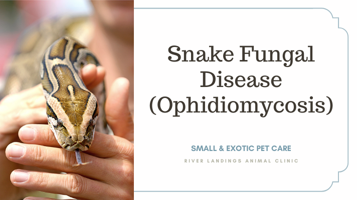
Snake Fungal Disease (Ophidiomycosis)

Cause of Snake Fungal Disease / Ophidiomycosis

Ophidiomycosis, formerly referred to as Snake Fungal Disease (SFD), is caused by the fungus Ophidiomyces ophiodiicola. This fungus is thought to be an emergent pathogen in North America and poses a significant threat to snake health and population sustainability. To date, O. ophiodiicola has been documented in over 15 types of wild and captive snakes.

Significance of Snake Fungal Disease / Ophidiomycosis

Ophidiomycosis is an emerging disease that affects wild and captive snakes across North America. The incidence of Ophidiomycosis has steadily increased over the last few years, specifically documented in California, Oklahoma, Puerto Rico, and Idaho.

Species Affected by Snake Fungal Disease / Ophidiomycosis

Ophidiomycosis can infect wild or captive snakes and is the predominant cause of skin infections in wild snakes. It has been documented in over 15 types of wild and captive snakes. Species confirmed with diagnosis of Ophidiomycosis include— the eastern indigo snake, the northern water snake, the eastern racer, the rat snake, the timber rattlesnake, the massasauga, the pygmy rattlesnake, garter snakes, cottonmouth snakes, the milk snake, queensnake, and eastern fox snake. The fungus causing Ophidiomycosis is not known to affect humans.

Distribution of Snake Fungal Disease / Ophidiomycosis

As of November 2019, the fungus O. ophiodiicola has been found in at least 38 states, including recent findings in California, Idaho, Oklahoma. It is also present in one U.S. territory (Puerto Rico) and one Canadian Providence (Ontario). In some species (i.e., Eastern Massasauga) the mortality rate may be over 90% in the Eastern US.

Transmission of Snake Fungal Disease / Ophidiomycosis

At this time, it is not well understood how the fungus causing Ophidiomycosis is spread. It is likely that the pathogen is shed into the environment by infected snakes and spread from the environment to other snakes (i.e., snakes that share dens). O. ophiodiicola may be spread via vertical transmission (i.e., from dam to offspring). There is no evidence of horizontal transmission (i.e., snake-to-snake transmission) of O. ophiodiicola. Host and environmental factors that lead to disease remain largely unknown. For example, it is thought that Timber rattlesnake populations are at higher risk of Ophidiomycosis in years with higher rainfall.

Clinical Signs of Snake Fungal Disease / Ophidiomycosis

Clinical signs of SFD and the severity of the disease may vary by species. The most common clinical signs include accelerated ecdysis cycles, flaking and crusting of the scales/epidermis, displaced or discolored scales, granulomas, nodules, swelling or disfiguration of infected tissues. The disease may progress internally (via the eyes, throat, and/or lungs) and cause eye infections or pneumonia.

Diagnosis of Snake Fungal Disease / Ophidiomycosis

Ophidiomycosis is diagnosed by identification of the classic clinical signs in combination with either molecular detection (PCR or qPCR) of the fungus, histopathology, and/or a positive fungal culture.

It must be noted that there may be difficulty in sampling for and detecting (by PCR/qPCR) O. ophiodiicola by skin/epidermal swabs (i.e., low DNA quantity on the skin or the fungi are deep within the epidermis). Therefore, hesitation should be taken in assigning causation to O. ophiodiicola to animals with skin lesions that are qPCR negative in the presence of skin lesions. There are numerous causes of skin lesions in wild snakes (i.e., trauma, another pathogen causing skin lesions, etc). Conversely, the absence of clinical signs of ophidiomycosis is more reliable to eliminate a diagnosis of apparent ophidiomycosis.

Treatment of Snake Fungal Disease / Ophidiomycosis

Treatment with an antifungal, Terbinafine, either by a slow-release, long-lasting implant or nebulization has been found to reach therapeutic levels in cottonmouth snakes; methods and doses for treatment are thus based on these findings. Other treatment options include thermal and nutritional supportive therapy.

Management of Snake Fungal Disease / Ophidiomycosis

Ophidiomycosis appears to be negatively impacting several species and populations of snakes across North America. However, its long-term impact remains unknown. It is also very difficult to assess changing snake populations because of the solitary nature of many snakes and, importantly, the absence of long-term monitoring data for most species of snake. Any sightings of snakes with suspected SFD infection should be reported to the Northeast Wildlife Disease Cooperative and/or DTT Herp Disease Alert System for further investigation. Wildlife specialists, veterinarians, and any other individuals who routinely come in contact with wild snakes should keep gear, snake holding containers, and clothes clean and disinfected to prevent spreading the fungus to other snakes.


Hear From Us Again

Don't forget to subscribe to our email newsletter for more recipes, articles, and clinic updates delivered straight to your e-mail inbox.

Related Categories:

Small Animal Care