Diabetes Ketoacidosis (DKA) - A Complication of Diabetes in Cats and Dogs

Unfortunately, as veterinarians, we are seeing an increased prevalence of diabetes mellitus in dogs and cats. This is likely due to the growing prevalence of obesity (secondary to inactive lifestyle, a high carbohydrate diet, lack of exercise, etc.). You're probably wondering if you just had a dog or cat diagnosed with diabetes mellitus—what do you do? First, we encourage you to take a look at these articles for an explanation of the disease:

This article will teach you about life-threatening complications that can occur as a result of the disease; specifically, a life-threatening condition called diabetes ketoacidosis (DKA) so that you know how to help prevent it.

What is DKA?

When diabetes goes undiagnosed or difficult to control or regulate, the complication of DKA can occur. DKA develops because the body is so lacking in insulin that the sugar can’t get into the cells — resulting in cell starvation. Cell starvation causes the body to start breaking down fat in an attempt to provide energy (or a fuel source) to the body. Unfortunately, these fat breakdown products, called “ketones,” are also poisonous to the body.

Symptoms of DKA

Clinical signs of DKA include the following:

  • Weakness

  • Not moving (in cats, hanging out by the water bowl)

  • Not eating or complete anorexia

  • Vomiting

  • Excessive thirst and urination (clear, dilute urine)

  • Large urinary clumps in the litter box (anything bigger than a tennis ball is abnormal)

  • Weight loss (most commonly over the back), despite an overweight body condition

  • Obesity

  • Flaky skin coat

  • Excessively dry or oily skin coat

  • Abnormal breath (typically a sweet “ketotic” odor)

  • Diarrhea

In severe cases DKA can also result in more significant signs:

  • Abnormal breathing pattern

  • Jaundice

  • Abdominal pain (sometimes due to the secondary problem of pancreatitis)

  • Tremors or seizures

  • Coma

  • Death

What can cause DKA?

When DKA occurs, it’s often triggered by an underlying medical problem such as an infection or metabolic (organ) problem. Some common problems that we see with DKA include the following:

  • Pancreatitis

  • Urinary tract infection

  • Chronic kidney failure

  • Endocrine diseases (e.g., hyperadrenocorticism [when the body makes too much steroid], or hyperthyroidism [an overactive thyroid gland])

  • Lung disease (such as pneumonia)

  • Heart disease (such as congestive heart failure)

  • Liver disease (such as fatty changes to the liver)

  • Cancer

Diagnosing DKA

While diagnosing DKA is simple, by looking at the blood sugar levels of dogs and cats and by measuring the presence of these fat breakdown products in the urine or blood, treatment can be costly (running between $3-5000). A battery of tests and diagnostics need to be performed, to look for underlying problems listed above, and treatment typically requires aggressive therapy and 24/7 hospitalization.

Treatment of DKA

Treatment, typically, is required for 3-7 days, and includes the following:

  • A special intravenous catheter called a “central line” (placed to aid in frequent blood draws)

  • Aggressive intravenous fluids

  • Electrolyte supplementation and monitoring

  • Blood sugar monitoring

  • A fast acting or ultra fast acting insulin, regular or Lispro, typically given intravenously or in the muscle

  • Blood pressure monitoring

  • Nutritional support (often in the form of a temporary feeding tube)

  • Anti-vomiting or anti-nausea medication

  • Antibiotics

  • Long-term blood sugar monitoring and a transition to a longer-acting insulin

Thankfully, with aggressive supportive care, many patients with DKA do well as long as pet parents are prepared for the long-term commitment (including twice-a-day insulin, frequent veterinary visits to monitor the blood sugar, and the ongoing costs of insulin, syringes, etc.).

Preventing DKA

By following your veterinarian’s guidelines and recommendations you can help regulate and control your pet’s diabetic state better and monitor your pet carefully for clinical signs. For example, if your pet is still excessively thirsty or urinating frequently despite insulin therapy, they are likely poorly controlled and need an adjustment of their insulin dose (of course, never adjust your pet’s insulin or medications without consulting your veterinarian).

When in doubt remember that the sooner you detect a problem in your dog or cat, the less expensive that problem is to treat. If you notice any clinical signs of diabetes mellitus or DKA, seek immediate veterinary attention. Most importantly, blood glucose curves (when a veterinarian measures your pet’s response to their insulin level) often need to be done multiple times per year (especially in the beginning stages of diabetes mellitus).

If you have any questions or concerns, you should always visit or call your veterinarian.

Related reading:


Hear From Us Again

Don't forget to subscribe to our email newsletter for more recipes, articles, and clinic updates delivered straight to your e-mail inbox.

Related Categories:

dog health, cat health

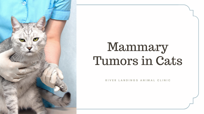
Can Cats Get Breast Cancer? Learn About Mammary Tumors in Cats

What is a mammary tumor (breast cancer)?

A mammary tumor develops as a result of abnormal replication of the cells that make up the breast tissue. Mammary tumors can be benign (non-cancerous) or malignant (cancerous). These two forms of the disease have different diagnostics, treatments, management, and prognosis. In cats, the vast majority of mammary tumors are malignant.

There are several different types of mammary tumors, with carcinomas being the most common. Carcinomas often arise from the tubules of the mammary glands, or other cells found in the mammary chain.

There are several other kinds of cancers that can affect the mammary glands, including osteosarcomas.

What causes this cancer?

The exact causes for the development of mammary tumors in cats are not fully understood. However, exposure to the female reproductive hormones, estrogen and progesterone, increases the risk for mammary cancers in cats. It is hypothesized that under the influence of hormones, the cells of the mammary gland enlarge, progress to a pre-cancerous state, and continue to grow until they transform into a carcinoma.

The incidence of these tumors is related to hormone status, specifically whether a cat is intact or has had an ovariohysterectomy (i.e., has been spayed). Sexually intact cats have a 7-fold increased risk for mammary tumors compared to spayed cats. Early spaying helps to prevent the development of these tumors. Cats that are spayed before 6 months of age have only a 9% percent risk of developing mammary tumors, whereas the risk increases to 14% in cats spayed between 7 and 12 months. Male cats rarely develop mammary tumors.

Age and breed also play a role in tumor development. Mammary cancers are seen mainly in middle-aged to older cats (around 10 years of age). Siamese cats appear to be predisposed and are typically younger when diagnosed. Obesity may play a role in tumor development as well.

What are the signs that my cat has this type of tumor?

The most common clinical sign of a mammary tumor is one (or more) palpable masses (nodules) underneath the skin of the abdomen. They may be next to or within the nipple and follow along the mammary chain. The size of the mass(es) and their appearance may vary, but they are usually firm and nodular. Occasionally the skin over the mass may ulcerate (open) and bleed, and the affected area may feel warm to the touch and become painful. Your cat may tend to lick and groom the area excessively, and a strong odor can result as the exposed tissue becomes infected.

If the tumor has metastasized (spread to other areas of the body), other signs may become evident. Your cat may generally feel unwell, eat less, become lethargic, and lose weight. If the lungs are involved, your cat may have problems breathing or develop a cough.

Multiple tumors are common at the time of diagnosis.

How is this type of tumor diagnosed?

These tumors are typically found as either singular or multiple nodules along your cat’s abdomen during a physical exam. Other procedures are then performed to determine the type of tumor and whether it is benign or malignant.

A common procedure to diagnose this tumor is a fine needle aspiration (FNA). FNA involves taking a small needle with a syringe and suctioning a sample of cells directly from the tumor and placing them on a microscope slide. A veterinary pathologist then examines the slide under a microscope.

In some cases, results from FNA may not be entirely clear and removal of the entire tumor may be necessary. Pieces of the tumor are then examined by a veterinary pathologist under a microscope. This is called histopathology. Histopathology is not only helpful to make a diagnosis but can indicate whether it is benign or malignant, and therefore how the tumor is likely to behave.

Given the risk of metastasis (spread to other areas of the body) with malignant mammary tumors, your veterinarian may also recommend staging (searching for potential spread to other locations in the body). This may include bloodwork, urinalysis, X-rays of the lungs, and possibly an abdominal ultrasound. Lymph nodes associated with the mammary glands may be sampled by FNA even if they seem normal.

How does this cancer typically progress?

The way this cancer progresses is entirely dependent on the type and size of the tumor(s), and whether metastasis has occurred. Larger tumors (greater than 2 cm) and those with evidence of spread have a poor prognosis. If the histopathology report shows tumor cells invading the local blood vessels, this also carries a poor prognosis. Detecting and treating these tumors when they are small and prior to metastasis provides your cat with the best chance of long-term control. Tumors smaller than 2 cm carry the best prognosis.

What are the treatments for this type of tumor?

Surgery is by far the best treatment for cats with mammary tumors. Depending on whether there is a single mass present, or several masses a single chain mastectomy (removal of one side of the breast tissue) or double chain mastectomy (removal of both sides, with each mastectomy performed at a different time) will be performed. Depending on the tumor size and other factors, chemotherapy is typically pursued afterward, especially given the high metastatic rate in these tumors.


Hear From Us Again

Don't forget to subscribe to our email newsletter for more recipes, articles, and clinic updates delivered straight to your e-mail inbox.

Related Categories:

cat health

Road Safety and Traveling with Pets

A study provided by Volvo Car USA and The Harris Poll revealed that allowing pets to roam unrestrained on road trips led to significantly more unsafe driving behaviors, more time spent distracted, and increased stress on both the drivers and their pets.

Some of their findings include:

  • 4 in 10 millennials choose a weekend getaway with their dog than their partner

  • 32% of pet owners have left a dog at home because they felt their car was not safe enough

  • 77% of Americans say people don't take vehicular dog safety seriously enough

It’s well established by now that our pets are essential members of the family, but many pet owners aren’t keeping their safety in mind when driving with them too. Volvo’s report followed 15 drivers and their dogs for more than 30 hours on the road to examine how driving with an unrestrained pet affected driving behavior vs. when an owner used a restraint (pet seat belts, harnesses, crates, carriers, etc).

What Happens With Pets Allowed to Roam Freely

Unsafe driving behaviors more than doubled. 649 instances while unrestrained vs. 274 while restrained. This includes climbing on a driver’s lap or hanging their head out of the window.

Time spent with drivers distracted more than doubled. 3hrs 39 mins spent unrestrained vs. 1hr 39 restrained. This includes dogs jumping from seat to seat or taking the drivers’ eyes off of the road.

Stress on both drivers and pets increased. Heart rates were likely increased for both people and pets. Unrestrained dogs measured a heart rate 7 beats per minute faster. Drivers felt calmer when dogs were buckled in, with heart rates dropping as much as 28 and 34 beats per minute.

The study further illustrates the dangers to all passengers, human and canine, when pets are not restrained. While pets roaming around the car can be cute and convenient, it poses serious risk for both drivers and their pets, both in terms of causing distractions and increasing the chances of serious injury in the event of an accident.

How to assure a safer road trip with pets

  1. Don’t drive with your pet in the front seat. In the event of a collision, a dog or cat can be thrown from the seat into the windshield, even if restrained. Unless the passenger side airbag is disengaged, the airbag deployment can be dangerous to a small pet.

  2. Never drive with your pet on your lap. It is not only a serious distraction to driving, bu the pet can get caught under the steering wheel and cause and accident or can be projected forward in the event of a collision.

  3. Never drive with your pet unrestrained. In addition to being a distraction, an abrupt can cause the pet to fall quickly and cause soft tissue injury or worse. In the event of an accident, pets can jump from the vehicle and go into moving traffic and be hit by other vehicles or get lost in an unfamiliar area.

  4. Never allow your pet to lean out of a car window. Debris can be forced into their eyes and cause abrasions or punctures to the cornea / eye, which can result in blindness.

  5. Never leave your dog unattended in a vehicle. Depending on the breed, level of anxiety, and ambient temperature, some people may be tempted to keep their pet in the car while running a short errand. Even in cooler months, never leave your dog unattended in your vehicle, no matter how short of a period, to avoid extreme temperatures and heat stroke / hypothermia.

With these tips in mind, we wish everyone safe, future road trips with their pets!


Hear From Us Again

Don't forget to subscribe to our email newsletter for more recipes, articles, and clinic updates delivered straight to your e-mail inbox.

Related Categories:
dog health, cat health

How can you tell when a cat is in pain?

Assessing pain is a complicated challenge, especially in cats. Pain has two primary components: the sensory aspect (intensity, location and duration) and the affective aspect (emotional toll).

Because pain assessment is somewhat subjective, veterinarians constantly try to create tools that make this process more objective. For validity, any pain measuring tool should take into consideration both characteristics: the sensory and the affective.

Signs of pain in cats

A British study was recently conducted in order to reach a consensus about criteria when evaluating pain in cats. Ultimately, 25 signs were considered to be reliable and sensitive for indicating pain in cats, across a range of different clinical conditions:

Top 5 Signs:

  • Appetite decrease

  • Avoiding bright areas

  • Growling

  • Groaning

  • Eyes closed

Other signs included: Lameness, difficulty jumping, abnormal gait, reluctant to move, reaction to touch, withdrawing/hiding, absence of grooming, playing less, overall activity decrease, less rubbing toward people, general mood, temperament, hunched up posture, shifting of weight, licking a particular body region, lower head posture, eyelids tightly shut, change in form of feeding behavior, straining to urinate, tail flicking

The top 5 signs are indicative of severe pain. Behavioral changes, such as irritability, tend to be seen with more long-term pain. The other signs can be observed with less intense pain. All of these signs cover both the sensorial and the emotional aspects of pain.

What if you see these signs of pain in your cat?

Cat owners should be aware of these signs. It is easy to mistakenly attribute behavioral changes, such as absence of grooming or playing less, as signs of aging; they can actually be signs of pain.

Remember, the presence of any single one of these 25 signs means pain. If you see any of these signs in your cat, see your veterinarian right away. Also remember that the absence of a sign does not mean your cat is no pain.

These signs may help both vets and cat guardians better assess the pain status of cats in their care.

While it can be fairly easy to recognize severe pain, it is much more difficult to detect low grade pain. The criteria above are a great start. Hopefully, this research will spark more studies to help us assess mild pain in cats as well to ensure their well-being.

If you have any questions or concerns, you should always visit or call your veterinarian— they are your best resource to ensure the health and well-being of your pets.


Hear From Us Again

Don't forget to subscribe to our email newsletter for more recipes, articles, and clinic updates delivered straight to your e-mail inbox.

Related Categories:

cat health

5 Signs Your Pet Has an Eye Emergency

5 Signs Your Pet Has an Eye Emergency

It's not uncommon for a veterinary clinic to field plenty of questions concerning pet eye problems on the daily. “Watchful waiting” is advised with some minor complaints, but never with eyes.

A non-vet pet owner cannot assess an eye problem’s severity, and a description over the phone just doesn’t cut it. In other words, when someone calls in and says their pet’s eye “looks funny,” it’s time for a trip to the vet.

Your vet may even direct you to a board-certified veterinary ophthalmologist for immediate assessment if the emergency requires it.

Red, inflamed or swollen eyes? Each of these symptoms can be caused by different ailments.

1. Red Eye

The white of the eye should be pristine white, with the odd lazy blood vessel meandering across the surface. If you’re not sure what this looks like, check out your own eye in the mirror.

Red eyes are not normal.

Gently lift the upper eyelid to check, and you’ll see anything from a rosy pink to a livid red. As a rule of thumb, the angrier the eye looks, the more urgently it needs checking.

Causes of red-eye range from irritation and infection to a condition called glaucoma.

Glaucoma: Pressure builds within the eye, like blowing too much air into a balloon. The most common cause is a problem within the eye and is often breed-related (Basset Hounds, Cocker Spaniels, terriers and northern breeds are at greatest risk). The pressure can damage the retina and cause blindness, so swift action is essential.

Conjunctivitis: Infection causes reddening of the eye. Sometimes the problem can be self-limiting, but — especially if there is a sticky yellow-green discharge, too — please see a vet.

2. Yellow-Green Discharge

It’s normal to have a white gloop in the corner of an eye first thing in the morning. Just wipe this away with a clean, damp cotton towel.

Also, rust-colored gloop is fine. This is a normal gloop that’s been exposed to the air for a while and oxidized (like when you cut an apple in half and it goes brown).

What isn’t normal is a thick, yellow-green discharge from one or both eyes. This is commonly a sign of infection.

Have the vet check the eye because some infections need antibiotics, while others can occur as a complication of another problem that needs attention.

3. Swollen Eye

If there’s something odd about your pet’s face, compare one eyelid with the other to see if one side is swollen. Eyelid swelling can be the result of an allergy, trauma, or infection. It’s best to seek vet attention because the eyeball needs checking to make sure it wasn’t damaged.

4. Dull Eye

Is the eye lackluster? A normal eye is bright, and you can see reflections on the surface— but sometimes the surface is dull and reflections aren’t clear, or those images are broken up or haphazard.

The most common reasons for this are either a dry eye or a corneal ulcer:

Dry eye: Our eyes are kept comfortable by the production of tear fluid. A pet with dry-eye fails to produce enough tear fluid, which leads to the surface drying out. One consequence is a dull surface, and another is the eye tries to protect itself by producing a thick, glue-like discharge. In the long term, scar tissue forms, impairing the vision.

Corneal ulcer: This is like a burst blister on the surface of the eye. In some cases, it heals on its own, but other times it can be dangerous and cause perforation of the eye.

5. Closed Eye

A closed eye is painful: Just think of the last time you had grit in your eye.

The pain might be due to a corneal ulcer, a knock to the eye, or a foreign body — anything from dust to grass particles or even a twig. When our pets are sniffing around, it’s not uncommon to get something lodged behind the eyelids.

Your vet will put drops of local anesthetic into the eye to get a better look and remove the object.


5 Most Common Eye Emergencies (in Dogs)

Here, we go into more detail about some of the top ophthalmologic emergencies we see with dogs in general practice.

1. Corneal Ulcers

Corneal ulcers are the most common acute eye problems seen. (This goes for cats as well.) The cornea is the outer “skin” of the eye; an ulcer occurs if this layer has been damaged.

Corneal ulcers are often traumatic in origin, although certain diseases of the cornea can result in an ulcer. Trauma to the cornea can occur with a scratch from a bush, a stick or even another critter.

Treatment involves early diagnosis of the severity of the ulcer and the administration of appropriate eye medications.

More serious ulcers may require surgery, and frequent re-checks are needed to ensure the cornea is healing nicely. When a corneal ulcer goes from bad to worse, the cornea can actually rupture — this is a true emergency indeed.

2. Proptosis

Proptosis occurs when an eye literally bulges out of the socket and the eyelids entrap the globe. This occurs most frequently in brachycephalic breeds and is a true emergency.

Even with immediate care, the dog may lose the eye depending on the extent of trauma sustained by the extraocular muscles, nerves, and blood supply.

Many of these eyes can be replaced and vision saved in about 20% of dogs, but only if you act immediately.

3. Corneal Laceration

Corneal laceration occurs when there is a complete tear through the cornea. The most common offender from a cat claw.

A sharp object, like a stick, can also puncture the cornea. The dog is almost always holding the eye completely shut and is in significant pain. Again, immediate surgery may save the eye.

Get to the vet. There is a very small window of opportunity when it comes to repairing these eye injuries.

4. Lens Luxation

Lens luxation is over-represented in certain breeds like the Russell Terrier and other terriers. In these dogs, due to a genetic disorder, the lens can spontaneously luxate, or become dislocated. In other breeds, causes may vary — head trauma is one example. This is a difficult diagnosis and may require a visit to an ophthalmologist.

Compared with the normal eye, a lens luxation can look like a very dilated pupil or a blue or whitish eye. Removing the lens can save the eye and save the dog from pain and total blindness.

5. Acute Glaucoma

Acute glaucoma looks like a discolored, a “red” or an inflamed eye. There may be discharge and painful blinking (called blepharospasm).

Glaucoma occurs when, for whatever reason, the pressure in the eye elevates, leading to pain, secondary changes, and blindness. The condition is usually obvious in just one eye, but both eyes are at risk.

Again, this is over-represented in certain breeds like the Cocker Spaniel, Basset Hound, Shih Tzu, Great Dane, and northern breeds.
As with every other eye problem discussed here, acute glaucoma should be assessed and treated immediately. Both systemic (oral) and ophthalmic drugs are used. Get an opinion from a veterinary ophthalmologist to help you and your primary care vet manage the case.

Don’t mess around with the eyes. Ophthalmic problems need early diagnosis and treatment.


Hear From Us Again

Don't forget to subscribe to our email newsletter for more recipes, articles, and clinic updates delivered straight to your e-mail inbox.

Related Categories:

DOG HEALTH

CAT HEALTH